
Check out some of my projects
Biomedical Communicator | Translating clinical and biomedical research across surgical and medical disciplines
Location
University Hospital Limerick, Limerick
University of Limerick
Biomedical Communicator | Translating clinical and biomedical research across surgical and medical disciplines
University Hospital Limerick, Limerick
University of Limerick
Data Analysis
AI Assisted CT Analysis of Fat Distribution in the Living Abdomen
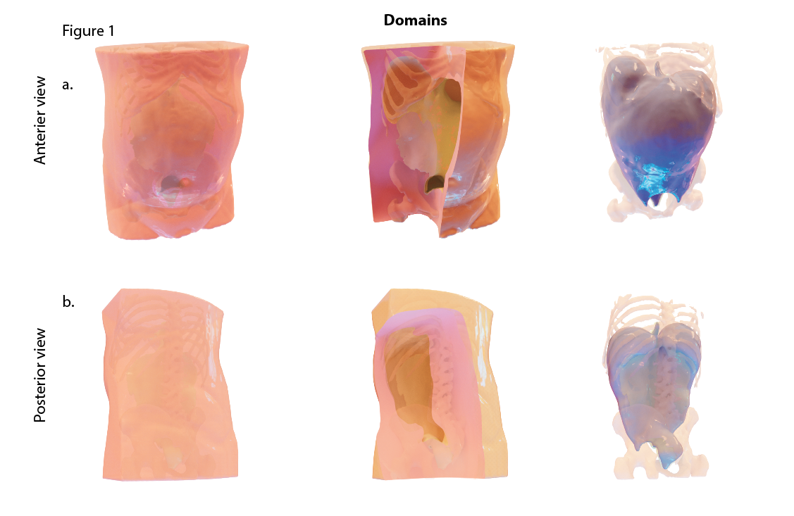
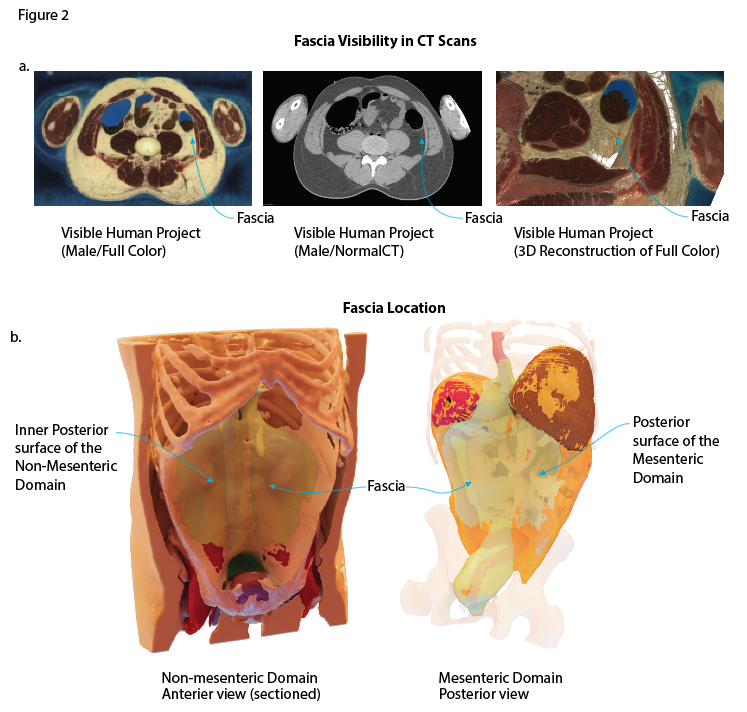
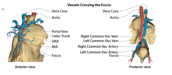
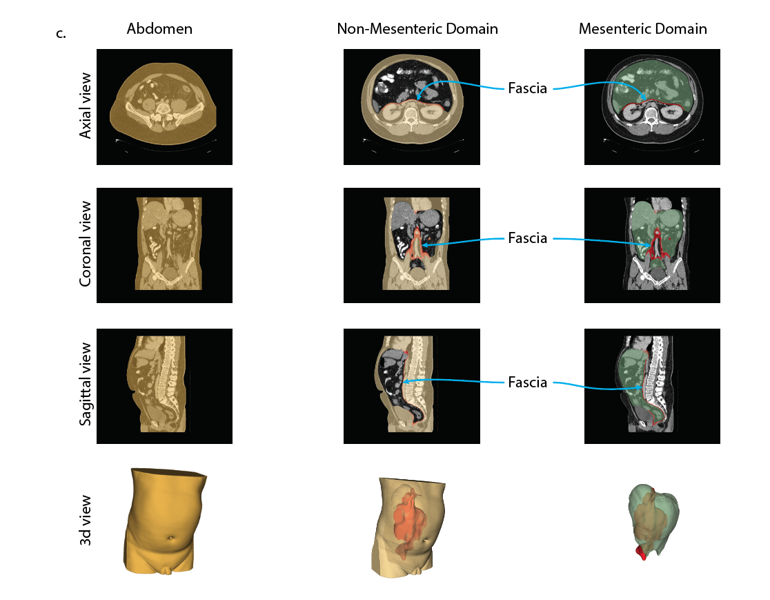
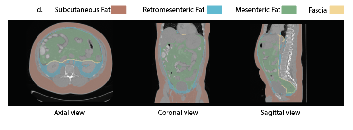
This project measures abdominal fat on routine CT imaging using an anatomical framework rather than an imaging convention. Fat is traditionally described as visceral or subcutaneous, a division based largely on appearance on the scan. The mesenteric model instead organises the abdomen into two domains, mesenteric and non-mesenteric, separated by a continuous fascia. Each fat depot can then be assigned to the domain in which it sits. By applying this framework across a large cohort of adult scans, the project aims to describe what normal fat distribution looks like and how it differs between men and women and across the lifespan.
 Open full-size image (new tab)
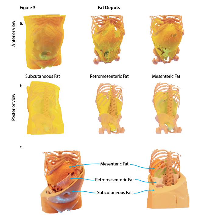
Where fat is stored in the body matters as much as how much there is. Fat within the abdomen is closely linked to metabolic and cardiovascular disease, yet the categories used to measure it have rarely been grounded in anatomy. Defining depots by the domain they occupy provides a consistent, reproducible way to compare individuals and to study how fat distribution relates to health and disease.
 Open full-size image (new tab)
 Open full-size image (new tab)
CT scans are processed through an automated segmentation pipeline built on AI tools, including nnU-Net, TotalSegmentator and MONAI Label. The models identify the abdominal domains, the fascia between them and the individual fat depots, including mesenteric, retromesenteric and subcutaneous fat. Trained annotators review and correct outputs to ensure anatomical accuracy. Volumes are then calculated for each depot and linked with basic demographic information.
 Open full-size image (new tab)
The project involves building and maintaining a structured research dataset that combines imaging derived measurements with clinical records. Processing runs on cloud GPU infrastructure, and quality control checks are applied before any analysis. Statistical analysis and figures are produced in R, using reproducible workflows so that every table and figure can be regenerated directly from the source data. Three dimensional reconstructions of domains, fascia and depots are used to communicate the findings visually.
 Open full-size image (new tab)
 Open full-size image (new tab)
The work is currently being prepared for publication. Future phases will link fat distribution with clinical outcomes and extend the approach to disease cohorts, building on the wider mesentery research programme and the AI medical imaging work described elsewhere on this site.